Cancer Detection with Image Processing and Artificial Intelligence
Cancer Detection with Image Processing and Artificial Intelligence
Introduction
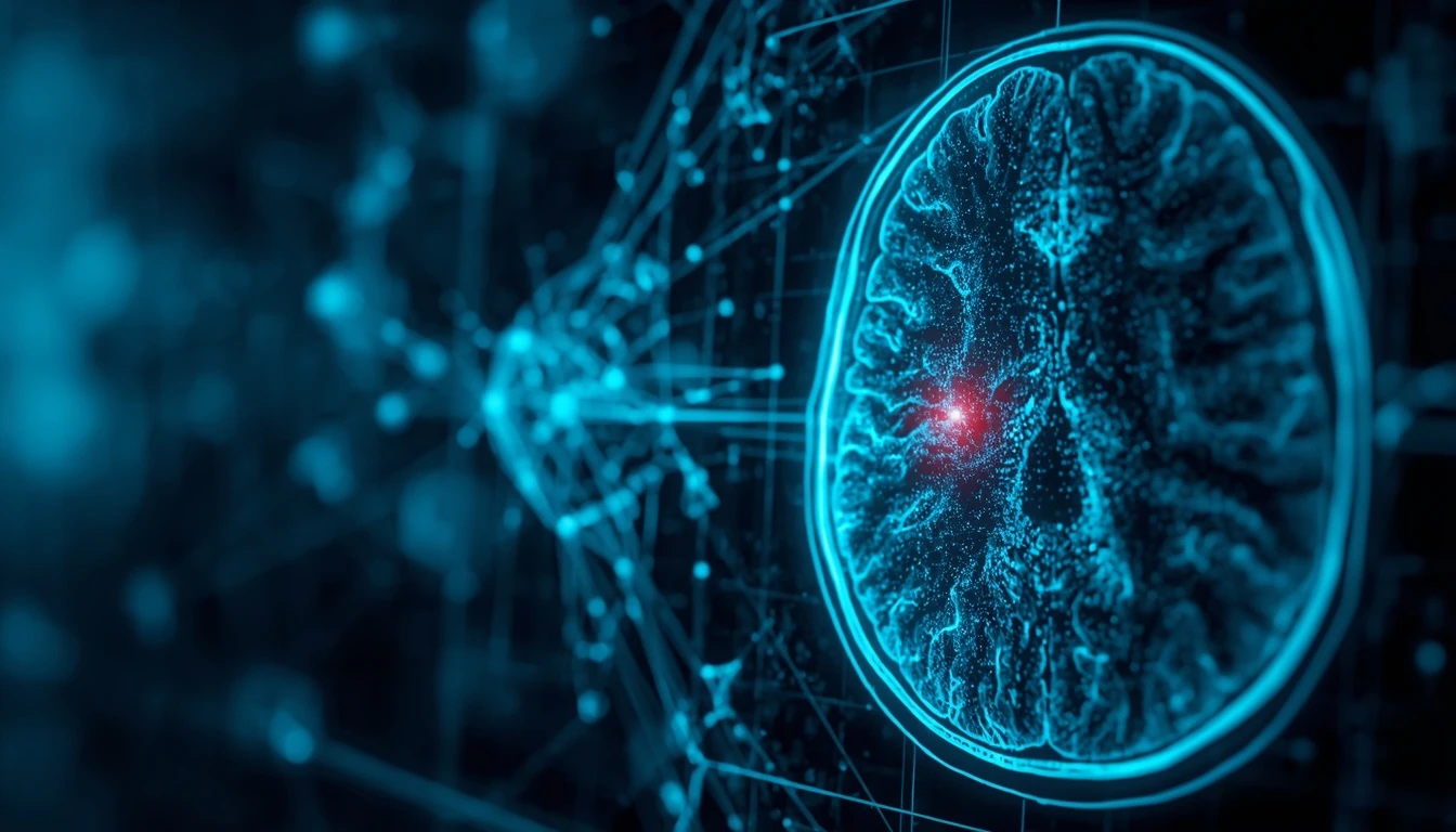
Early cancer detection is one of the major challenges in modern medicine. Doctors use various imaging techniques, including MRI, CT scans, mammography, ultrasound, and pathology images, to identify tumors and suspicious lesions. As the volume of medical images continues to grow, reviewing all of these images accurately can require significant time and expertise.
This is where image processing and artificial intelligence can provide valuable support. AI algorithms can analyze large volumes of medical images and identify patterns that may be difficult to recognize consistently through human inspection alone. The National Cancer Institute highlights AI research in areas such as cancer screening, diagnosis, and medical image analysis.
How Does Medical Image Processing Help Detect Cancer?
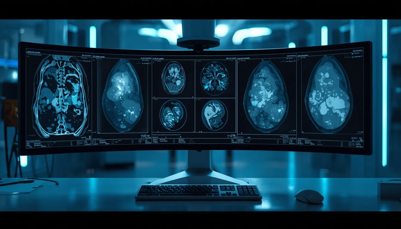
Medical image processing uses computational techniques to extract useful information from images of the human body. In an intelligent system, an image can first be preprocessed to reduce noise and improve the quality of relevant information. AI algorithms can then identify, classify, or segment different regions within the image.
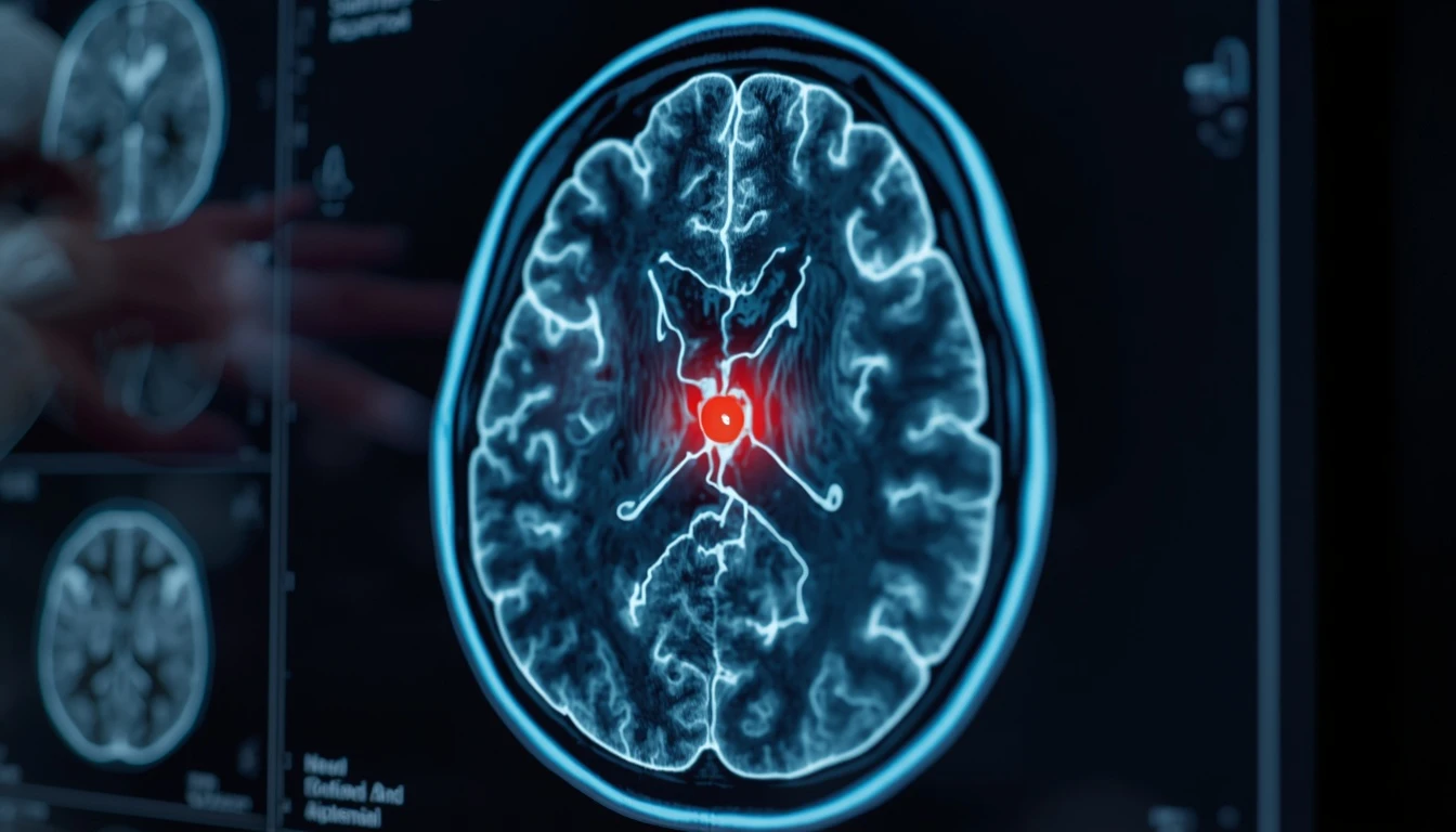
For example, an AI model can analyze an MRI scan and identify an area with characteristics associated with a tumor, or examine pathology images to highlight abnormal cellular structures for further review. Cancer research has explored these approaches for lesion detection, tumor measurement, and tissue analysis.
Pishgaman Lotus can contribute to the software and AI development side of such solutions by combining computer vision, machine learning, and software engineering. However, clinical use requires scientific validation and compliance with applicable medical and regulatory requirements.
The Role of Deep Learning in Tumor Detection
One of the key technologies used for medical image analysis is Deep Learning. Deep learning models use neural networks to learn patterns from large datasets and can be trained to distinguish between different types of medical images or tissue characteristics.
During training, an AI model can learn from medical images that have been reviewed and labeled by specialists. The model gradually learns visual patterns associated with healthy and abnormal tissue. After training and validation, it can analyze new images and highlight areas that require further examination.
NCI research has explored deep learning applications involving prostate MRI, pathology images, and cancer screening.
Cancer Detection in MRI and CT Images
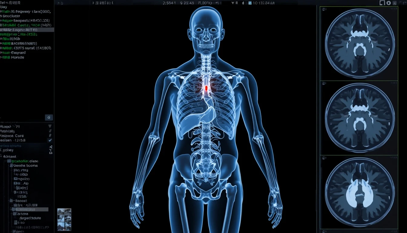
MRI and CT scans are important sources of data for AI-based medical imaging systems. Artificial intelligence can assist with identifying suspicious areas, separating tumors from surrounding tissue, and measuring the size or volume of lesions.
An important application is image segmentation. In this process, an algorithm attempts to distinguish a tumor or organ from surrounding structures. Such information can help measure tumors and monitor how they change over time.
Recent research has also explored AI-assisted analysis of whole-body images, where AI can help identify and segment organs and suspicious regions across large imaging datasets.
Artificial Intelligence in Breast Cancer Detection
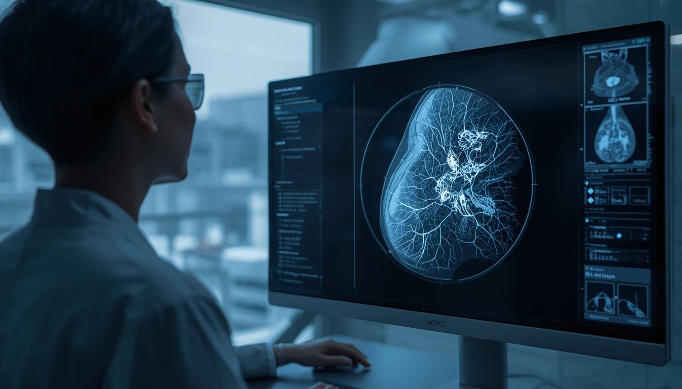
One of the areas where AI has been extensively studied is mammography and breast cancer detection. The large volume of mammographic images and the importance of identifying small abnormalities have encouraged the development of computer-assisted analysis tools.
AI systems can process mammograms rapidly and highlight suspicious regions for further review. Some research has also investigated whether mammographic images can be used to estimate future breast cancer risk.
The purpose of these systems is not necessarily to replace physicians. Instead, they can serve as supporting tools that make parts of the image-review process faster and more consistent.
Cancer Detection in Digital Pathology
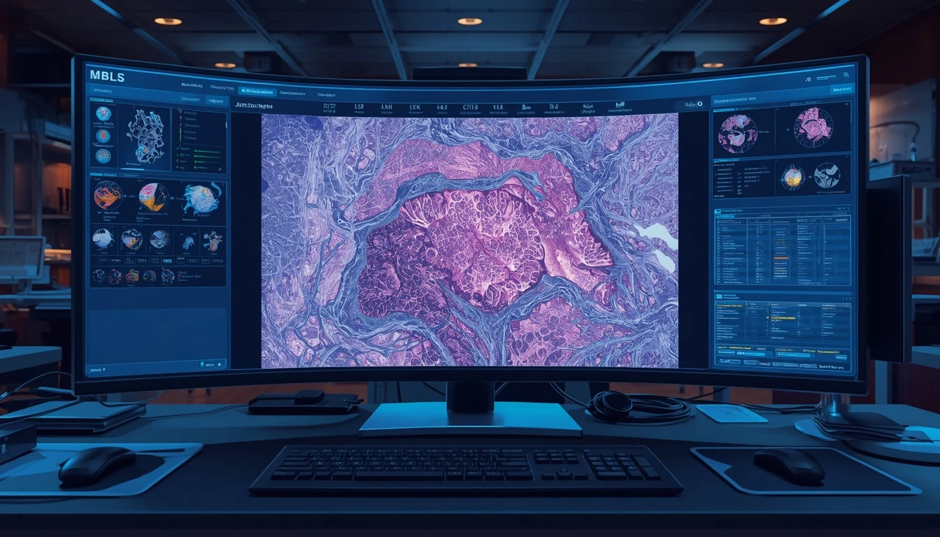
AI applications are not limited to MRI and CT scans. Digital images of tissue samples can also provide valuable data for computer vision models.
A pathology image may contain enormous numbers of cells and tissue structures. AI algorithms can analyze these images and identify patterns associated with abnormal cells or specific tumor characteristics.
NCI research has also explored the use of AI to analyze pathology images and investigate relationships between tumor appearance and molecular characteristics.
Benefits of AI for Cancer Image Analysis
One of the major advantages of AI is its ability to process large volumes of data quickly. An AI system can analyze numerous medical images using consistent computational processes and provide preliminary results for specialist review.
Reproducibility is another important factor. A properly designed and validated algorithm can perform the same analytical task using consistent criteria across a large dataset. AI may also help identify complex visual patterns that can be difficult or time-consuming for humans to assess.
For Pishgaman Lotus, this field represents an example of how artificial intelligence can transform complex visual data into structured and potentially useful information for specialized software systems.
Can Artificial Intelligence Replace Doctors?
Despite rapid technological progress, AI-based cancer imaging should still be used with professional oversight. Model performance can vary depending on the imaging technique, training data, patient population, and clinical context.
A 2025 NCI study comparing AI and radiologists found that AI was particularly useful for rapidly and consistently identifying and measuring localized tumors, while radiologists performed better in some aspects of assessing tumor extent. The study therefore described AI as an adjunct to radiologists rather than an independent replacement.
A more practical direction is therefore collaboration between physicians and AI systems. AI can analyze large volumes of imaging data and highlight patterns, while physicians combine those results with clinical history, expertise, and other medical information when making decisions.
Challenges of AI-Based Cancer Detection
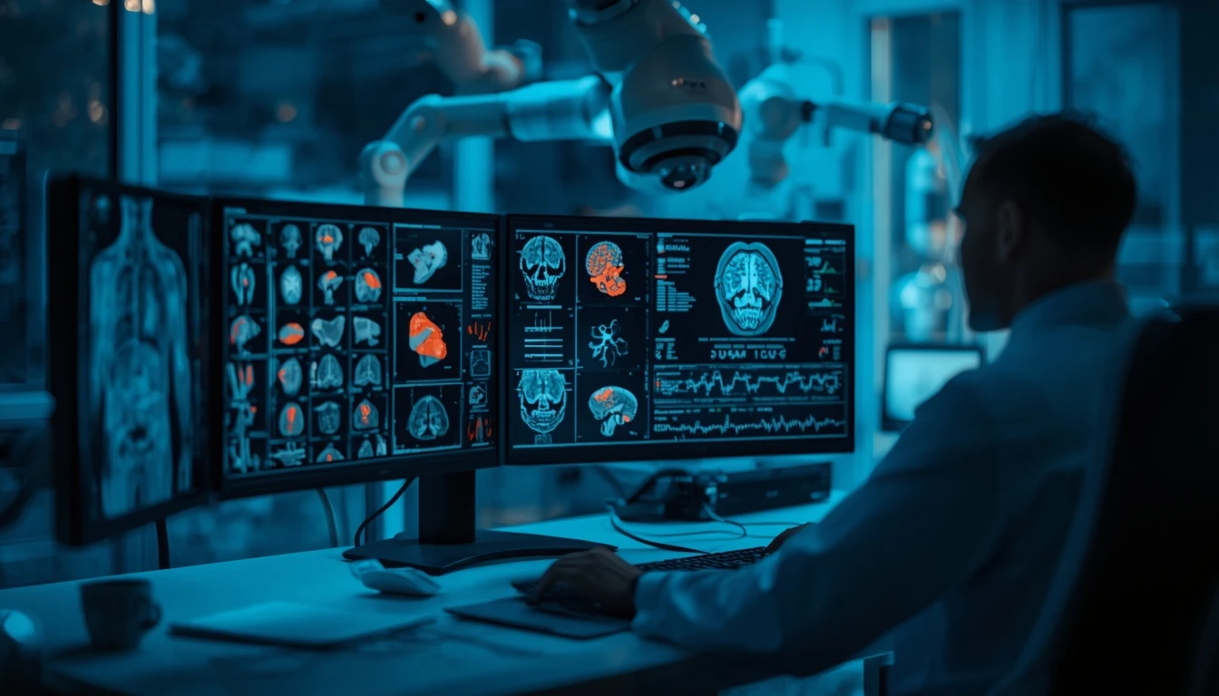
Developing an AI medical imaging system involves much more than building a machine learning model. Data quality, dataset diversity, accurate labeling, patient privacy, model validation, explainability, and clinical evaluation are all important considerations.
If training data do not adequately represent real-world populations, model performance may vary in other settings. In addition, some deep learning systems can be difficult to interpret, making it challenging to understand why a particular image was classified as suspicious. This is especially important in medicine, where clinical decisions need to be carefully evaluated.
For this reason, medical AI development requires rigorous testing, secure data management, scientific validation, and appropriate regulatory processes.
The Future of Cancer Detection with Image Processing
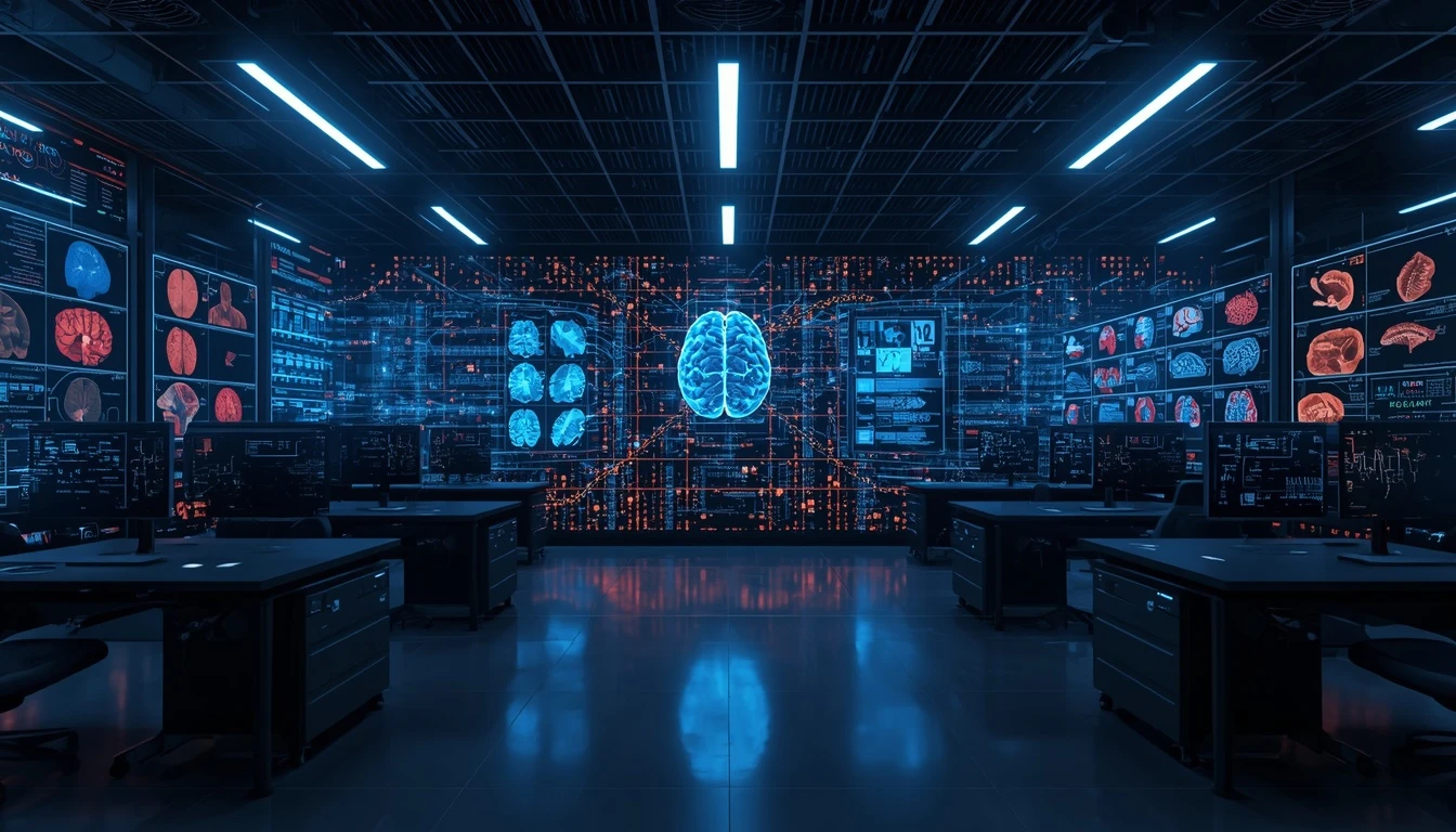
The future of AI-assisted cancer detection is likely to involve the integration of multiple types of medical data. Instead of analyzing a single image independently, future systems may combine imaging data with clinical information, pathology, and molecular characteristics.
Three-dimensional and whole-body imaging may also provide broader opportunities for detecting and tracking tumors. NCI research is already exploring AI-based approaches for identifying, segmenting, and monitoring tumors across different types of medical images.
In this field, Pishgaman Lotus can leverage its capabilities in artificial intelligence and software development to contribute to the design of technical infrastructures and specialized software solutions for data-intensive applications.
However, a research or software-based AI model should not automatically be considered a clinically approved diagnostic system. Clinical deployment requires appropriate scientific validation, regulatory approval, and medical oversight.
Conclusion
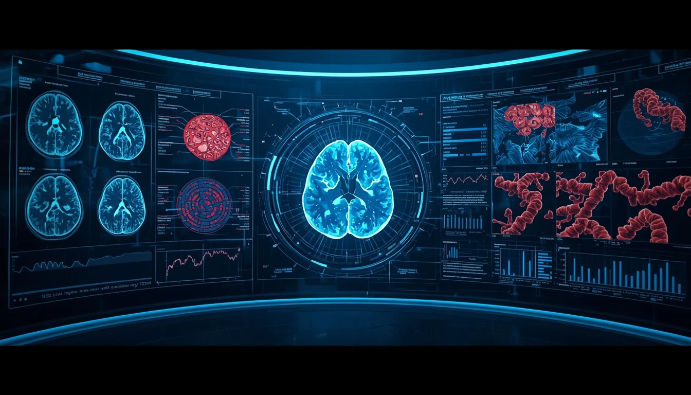
The combination of image processing and artificial intelligence represents an important direction in modern medical technology. From MRI and CT analysis to mammography and digital pathology, AI can help identify patterns, highlight suspicious regions, measure tumors, and process large volumes of medical images.
The real value of this technology, however, comes from combining AI capabilities with medical expertise, high-quality data, and rigorous validation. Rather than completely replacing physicians, the future of cancer imaging is more likely to focus on tools that enhance clinical expertise by providing faster and more structured analysis of complex medical data.
Pishgaman Lotus can contribute to this technological ecosystem through AI development, computer vision, and specialized software engineering, helping transform complex medical data into intelligent digital solutions.
Our articles:"Image Processing and Object Detection in Autonomous Vehicles"
Blog Tags
Similar Blogs
Image Processing and Object Detection in Autonomous Vehicles

Text-to-Video: The AI Revolution in Video Creation
Industrial Machine Vision: The Quality Control Revolution
Have special project to begin?
Contact us if you need a unique website for your special requirements, if you think having a mobile application help you reach your business’s goals or you still do not recognize which product can help you implement your ideas. Lotus Pioneers accompany you to develop your business through consulting and by designing special products.